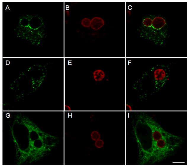
Figure 4.
Localization of Act1 in C. pneumoniae infected cells. C. pneumoniae infected cells were transfected with FLAG-tagged Act1 at 45 hrs p.i. and cell cultures were fixed with methanol and stained with primary and secondary antibodies after an additional 19 hrs of incubation. (A) Act1 is associated with inclusion membrane of C. pneumoniae (green). (D) Association of Act1 was not detected with C. trachomatis inclusions in cells transfected with FLAG-tagged Act1. (G) Control experiment using C. pneumoniae infected cells transfected with 3xFLAG-CT847. Localization of chlamydial inclusions is depicted by staining with Chlamydia species-specific rabbit antiserum (B, E and H). Composite images (C, F and I). Scale bar = 10 μm.

