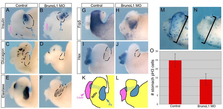
Figure 3. BrunoL1 is essential for endodermal cell differentiation.
(A,B) Expression of insulin was normal in the pancreas of brunol1 morphants (n=15). (C,D) Expression of glucagon in the stomach/duodenum is reduced (n=23). Similar results were found for somatostatin (n=20). (E,F) elastase expression was reduced (n=16). (G,H) Similarly, there was reduced expression of the general stomach marker, frp5 (n=14). (I,J) Liver development was normal as assessed by expression of hex (n=12). (K,L) Trace drawings of panels G and H. The pancreas is outlined in panels A–D, F, and I–J. (M) Control NF41 whole gut stained for phosphohistone H3 to mark proliferating cells. (N) Whole gut from brunol1 morphant stained for phosphohistone H3. Note the large decrease in proliferating cells, especially apparent in the stomach and duodenum. (O) Average number of proliferating cells within the stomach of 10 different whole guts from control and morpholino injected. (Error bars represent standard deviation.) We only counted the number of cells in the stomach/duodenal area, even though the morpholino was targeted to a larger region, including more posterior endoderm. To ensure that equivalent areas were compared we counted cells in control and morpholino samples within the same defined total area. A reduction in pH3 staining was also seen in the other targeted regions, but for quantification we only focused on the stomach/duodenal region. Areas not targeted by the morpholino (i.e. no fluorescence) showed normal pH3 staining.

