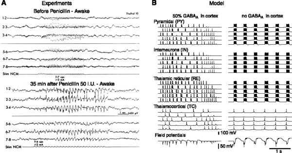
FIG. 13.
The 3-Hz spike-and-wave discharges obtained by altering cortical inhibition. A: spike-and-wave discharges following diffuse application of penicillin to the cortex of cats. Top: stimulation of nucleus centralis medialis (NCM; 7 Hz) induced a recruiting response in the cortex. Bottom: after diffuse application of a diluted solution of penicillin to the cortex (50 IU/hemisphere) in the same animal, 4-Hz stimulation of NCM elicited bilaterally synchronous spike-and-wave activity. Similar spike-and-wave discharges also occurred spontaneously. [Modified from Gloor et al. (130).] B: computational model of the transformation of spindle oscillations into ~3 Hz spike-and-wave by reducing cortical inhibition. A thalamocortical network of 400 neurons was simulated, and 5 cells of each type are shown from top to bottom. The last traces show the field potentials generated by the array of pyramidal neurons. Left: 50% decrease of GABAA-mediated inhibition in cortical cells. The oscillation begins like a spindle, but strong burst discharges appear after a few cycles, leading to large-amplitude negative spikes followed by slow positive waves in the field potentials. Right: fully developed spike-and-wave oscillations following suppression of GABAA-mediated inhibition in cortical cells. All cells had prolonged, in-phase discharges, separated by long periods of silence, at a frequency of ~2 Hz. GABAB currents were maximally activated in TC and PY cells during the periods of silence. Field potentials displayed spike-and-wave complexes. Thalamic inhibition was intact in A and B. [Modified from Destexhe (86).]

