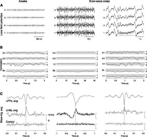
FIG. 17.
Spatial and temporal coherence of wake and sleep oscillations. A: bipolar local field potential (LFP) recordings at 4 equidistant sites (1 mm interelectrode distance) in suprasylvian cortex of cats during natural wake (left) and slow-wave sleep states (middle; note different time scale). Awake periods consisted in low-amplitude fast (20–60 Hz) activity while slow-wave sleep was dominated by waves of slower frequency (0.5–4 Hz) and larger amplitude. Right panel shows a period of slow-wave sleep with higher magnification, in which a brief episode of fast oscillations was apparent (dotted line). B: correlations between different sites, calculated in moving time windows. Correlations were fluctuating between high and low values between neighboring sites during fast oscillations (left), but rarely attained high values for distant sites (1–4; “Sh.” indicates the control correlation obtained between electrode 1 and the same signal taken 20 s later). In contrast, correlations always stayed high during slow-wave sleep (middle). Episodes of fast oscillations during slow-wave sleep (right) revealed similar correlation patterns as during the wake state. C: wave-triggered averages between extracellularly recorded units and LFP activity. During the wake state, the negativity of fast oscillations was correlated with an increase of firing (left; “control” indicates randomly shuffled spikes). The negativity of slow-wave complexes was correlated with an increased firing preceded by a silence in the units (middle; note different time scale). During the brief episodes of fast oscillations of slow-wave sleep (right), the correspondence between units and LFP was similar as in wakefulness. [Modified from Destexhe et al. (101).]

