Figure 3.
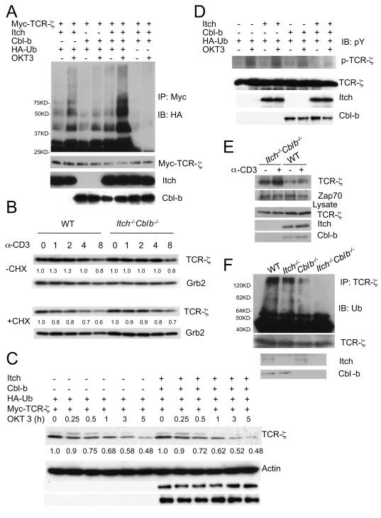
Itch and Cbl-b cooperate to promote TCR-ζubiquitination without degradation.
(A)Plasmids as indicated were transfected into Jurkat T cells and the cells were unstimulated or stimulated with OKT3 for 5 min, followed by the analysis of TCR-ζ ubiquitination. Molecular size markers were labeled. The Ub ladders are indicated. The membrane was reprobed with anti-Myc, and the lysates was immunoblotted with indicated antibodies.
(B) Primary T cells were stimulated with anti-CD3ε for different time intervals in the absence or the presence of cycloheximide and the cell lysates were immunoblotted with anti-TCR-ζ and the same membrane was reprobed with anti-Grb2. The intensity of the protein bands was quantified and the relative amount of TCR-ζ at each starting point of stimulation (0) was given an arbitrary unit of 1.0.
(C)Jurkat T cells were transfected with plasmids as indicated, and the cells were stimulated with OKT3 for different time intervals in the presence of cycloheximide and the lysates were immunoblotted with anti-Myc and the same membrane was reprobed with indicated antibodies. The intensity of TCR-ζ was quantified and the relative amount of TCR-ζ at time 0 was given an arbitrary unit of 1.0.
(D)Transfected Jurakt T cells were stimulated with anti-CD3 for 2 min and the lysates were analyzed with the indicated antibodies.
(E) T cells from WT or double-deficient mice were left unstimulated or stimulated with anti-CD3 for 5 min and the cell lysates were immunoprecipitated with anti-Zap-70. The immunoprecipitates were immunoblotted with anti-pY. The position of phosphorylated TCR-ζ was indicated. The same blot was reprobed with anti-Zap-70.
(F) Splenocytes from 8 week-old WT, Cblb−/−, Itch−/− or double-deficient mice were stimulated with anti-CD3 for 5 min and lysates were immunoprecipitated with anti-TCR-ζ, followed by immunoblotting with anti-Ub. . Molecular size markers are indicated at left.
See also Figure S3.

