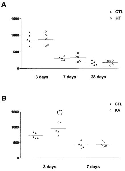
FIGURE 4.
Quantitative analysis of 5-bromo-2′-deoxyuridine (BrdU)-immunoreactive nuclei in the granule cell (GC) layer after (A) experimental febrile seizures (HT) lasting ~20 min, or (B) kainic acid (KA)-induced status epilepticus lasting ~120 min. A: Rats were injected with BrdU at 3, 7, or 28 days after the febrile seizures and perfused 48 h later. Data represent BrdU-positive nuclei counted in five sections (left and right hippocampus = 10 analyses per rat). The numbers of BrdU-labeled cells declined with age, due to the established developmental reduction in GC neurogenesis. However, no differences were found between HT and control animals at any age or time point. B: Rats were injected with BrdU at 3 or 7 days after KA-induced seizures and were perfused 48 h later. In contrast to the HT-induced seizures, KA-induced seizures significantly increased the number of BrdU-labeled cells at the 3-day time point (asterisk). The seizure-induced neurogenesis was transient and was no longer evident when rats were sacrificed 1 week after the seizures.

