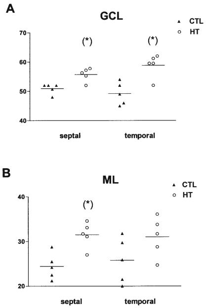
FIGURE 6.
Quantitative analysis of mossy fiber innervation of the granule cell (GCL, A) and molecular (ML, B) layers of the dentate gyrus in septal and temporal hippocampus (n = 5, each group). The hippocampal formation of naive 3-month-old rats (CTL) was compared with those from animals sustaining experimental febrile seizures early in life (HT). Sections were processed for Timm's stain as described in the methods, and all analyses were carried out without knowledge of treatment group. Significantly increased numbers of mossy fibers traversed the GCL in the HT animals (asterisks, A). More fibers also penetrated the ML in the septal (but not temporal) hippocampus (B). The y-axis denotes the numbers of fibers per linear millimeter (mm) of the suprapyramidal blade of the GCL.

