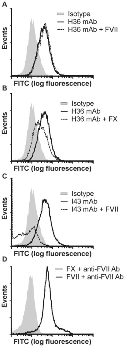
Figure 3. TF-specific staining of MDA MB231 cells with anti-TF mAbs H36 and I43.

(A) Tumor cells (2.2 × 10⁵ in 0.2 mL) were incubated with 0.5 μg of H36 in the absence (gray shading) or presence (dark line) of 6.5 μg human FVII then stained with FITC-labeled goat anti-mouse IgG. Staining was compared to cells treated with an IgG4 isotype control (dashed line). (B) Tumor cells were stained with H36 as in (A) in the absence (gray shading) or presence (dark line) of 10 μg of human FX. (C) Tumor cells were incubated with 0.7 μg of I43 in the absence (gray shading) or presence (dark line) of 6.5 μg human FVII then stained with FITC-labeled goat anti-mouse IgG. (D) Tumor cells were incubated with FVII (6.5 μg) (dark line) or FX (10 μg) (dashed line), and then with FVII antibody, followed by staining with FITC-labeled goat anti-mouse IgG. Positive staining of FVII treated cells verifies the ability of FVII to bind cell surface TF.
