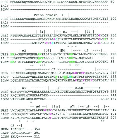
Figure 2.
Sequence alignment of Ure2p and structurally similar GSTs [PDB code 1A0F: E. coli GST (37); PDB code 1GNW: A. thaliana theta class GST (38)]. Secondary structural elements identified in the Ure2p(97–354) crystal structure are indicated above the sequence. Residues conserved in all sequences are indicated by an asterisk below the sequence. Catalytic residues are cyan. G-site (GSH-binding) residues are green. H-site (hydrophobic electrophile-binding) residues are violet. Underlined residues indicate residues contributed to a binding site by the second monomer of the dimer. The indicated residues in Ure2p have been selected by analogy to these similar GSTs.

