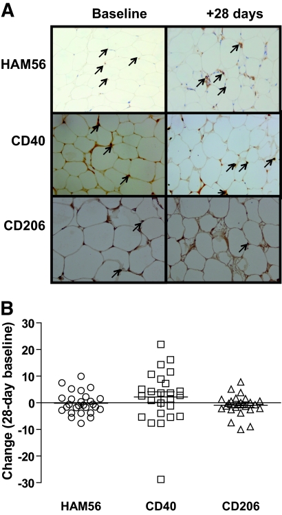
FIG. 2.
Immunohistochemistry staining of (A) macrophage-specific marker (HAM56), M1 marker (CD40), and M2 marker (CD206) phenotype macrophages in subcutaneous adipose tissue. Pictures are taken from paired representative slides, and positive cells are marked with a black arrow. B: Change in HAM56, CD40, CD206 positive cells from baseline in response to 28 days of overfeeding. (A high-quality digital representation of this figure is available in the online issue.)

