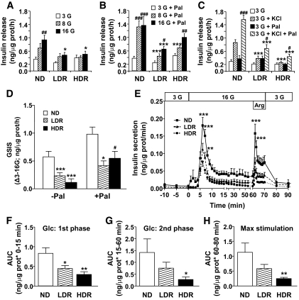
FIG. 3.
Isolated islets from DIO mice show defective glucose-, KCl-, and arginine-induced insulin release. Insulin secretion was measured in freshly isolated islets from normal diet (ND), LDR, and HDR mice. Group of 10 islets were incubated 1 h in KRBH at 3, 8, or 16 mmol/l glucose (G; A and D) ± 0.25 mmol/l palmitate/0.5% d-BSA (Pal; B and D) or 3 mmol/l glucose ± 35 mmol/l KCl (C) ± Pal (C). Means ± SE are of 14–16 determinations from islets of 21–22 animals per group in five (A, B, and D) and three (C) separate experiments. E–H: Insulin secretion by islets perifused at 3 mmol/l glucose (3G) or 16 mmol/l glucose (16G) with or without 10 mmol/l arginine (Arg). AUC for insulin over the first 15 min (F) and from 15–60 min (G) following glucose concentration increased from 3 to 16 mmol/l. AUC for maximal insulin secretion was determined from 60–80 min after arginine injection (H). Means ± SE are of six normal diet, seven LDR, and seven HDR. Insulin release in the media was determined after the 1-h incubation or at the different time points of the perifusion experiment. *P < 0.05, **P < 0.01, ***P < 0.001 normal diet versus LDR or HDR; #P < 0.05, ##P < 0.01, ###P < 0.001 versus 3 mmol/l glucose (A) plus palmitate (B) or plus KCl (C) or versus the value in absence of palmitate (D) for the same group (normal diet, LDR, HDR); one-way ANOVA–Bonferroni multiple comparison test.

