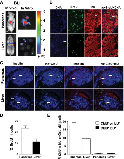
FIG. 5.
Islet graft β-cell proliferation/replication in the liver and pancreas as judged by BrdU-labeling and sequential CldU and IdU labeling. We transplanted 100 donor islets from luc+ FVB/N donors into the liver or pancreas of chimeric recipients. The day after islet transplantation, the recipients were given BrdU labeling or sequential CldU and IdU labeling. Islet grafts were harvested under the guidance of ex vivo BLI. Formalin-fixed graft tissues were stained for DNA, BrdU, and insulin; or CldU, IdU, and insulin. The proliferating β-cells were identified as BrdU+Insulin+. The replicating β-cells were identified with CldU+Insulin+ or IdU+Insulin+. β-cells from neogenesis were identified as CldU+IdU+Insulin+. Representatives of proliferating or replicating β-cells are indicated by arrows. A: In vivo and in vitro BLI of islet grafts. B: A representative staining pattern of BrdU-labeling of proliferating β-cells. C: A representative staining pattern of CldU and IdU-labeling of replicating β-cells. D: Mean ± SE of percentage of proliferating β-cells of 6 islet grafts in each group. E: Mean ± SE of percentage of β-cells from replication or neogenesis of 4 islet grafts in each group. (A high-quality digital representation of this figure is available in the online issue.)

