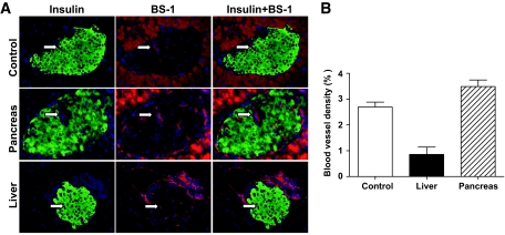
FIG. 6.
Graft islet revascularization in the liver and pancreas as judged by BS-1 staining. One hundred donor islets from luc+ FVB/N donors were transplanted into the liver or pancreas of chimeric recipients. Three weeks later, islet grafts were harvested under the guidance of ex vivo BLI. Formalin-fixed graft tissues were stained for DNA (blue), insulin (green), and BS-1 (red). Vascularization was identified with BS-1 staining (red) inside of insulin staining (green). A and B: A representative BS-1 staining pattern of control islets and islet grafts in the liver and pancreas (A); mean ± SE of percentage of vascular density of 4 islet grafts in each group (B). (A high-quality digital representation of this figure is available in the online issue.)

