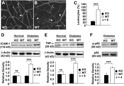
FIG. 2.
Analysis of retinal inflammation in conditional VEGF KO mice 2 months after inducing diabetes. A and B: FITC-conjugated ConA staining for adherent leukocytes (arrows) in retinal microvasculatures of diabetic conditional VEGF KO mice and WT controls. Scale bar represents 100 μm. C: Quantification of adherent leukocytes in retinal vasculatures of diabetic conditional VEGF KO mice and WT controls. D and E: Immunoblotting analysis of ICAM-1 (D) and TNF-α (E) expression in conditional VEGF KO mice. F: Immunoblotting analysis of NF-κB p65 phosphorylation in diabetic retinas of conditional VEGF KO mice. Error bar: SEM. ***P < 0.001. Loss of Müller cell-derived VEGF caused a significant reduction in the number of adherent leukocytes, expression of ICAM-1 and TNF-α, and phosphorylated NF-κB p65 in diabetic retinas. ns, not significant.

