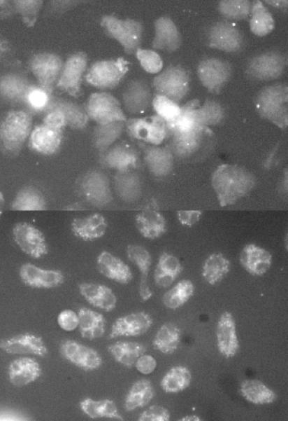
Figure 5.

Example images of degranulation sac formation in a field of cells stimulated with anti-IgE Ab (0.5 μg/ml) in the presence of vehicle control (DMSO) or Ro-31-8220 at 3 μM (cells pre-incubated with drug for 10 minutes prior to stimulation. Panel A; anti-IgE Ab with DMSO, image at ca. 10 minutes. Panel B; anti-IgE Ab with Ro-31-8220, image at ca. 10 minutes.
