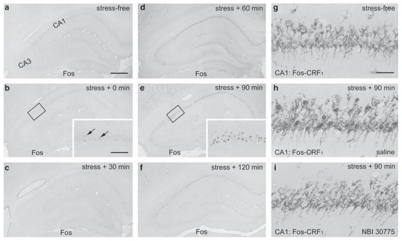
Figure 7.

Restraint stress induces Fos expression in adult hippocampal CA1, which can be blocked by prior infusion of CRF1 antagonists. (a–f) Time course of Fos expression in adult hippocampus induced by 30-min restraint stress. (b, c) Rare Fos expressing neurons are apparent in the CA1 pyramidal cell layer immediately and 30 min after the termination of the stress (arrows in inset, b). (d, e) Strong Fos expression is detected in CA1 60 and 90 min after the stress. At these time-points, Fos is also expressed to a lesser extent in CA3. (g–i) CA1 neurons double-labeled for Fos and CRH receptor CRF1: Fos is not expressed in CRF1-expressing neurons of control animals implanted with cannula 6 days earlier (g). Stress-induced Fos expression (blue immunoreaction product) is co-localized with CRF1 (brown) and is not abolished by saline pre-administration through the preimplanted cannula (h, see Figure 1 for schedule). Prior infusion of the CRF1 antagonist NBI 30775 blocks restraint-induced Fos expression in CA1 (i). Scale bar = 720 μm (a–f), 180 μm (insets in b, c, e) and 60 μm (g–i).
