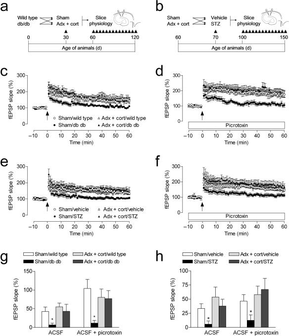
Figure 2. Lowering corticosterone levels regulates synaptic plasticity in diabetic rodents.
(a), Design for studies in Type 2 diabetic mice. (b), Design for studies in Type 1 diabetic rats. Adrenalectomized animals received corticosterone replacement via the drinking water (25μg ml–1 in 0.9% saline). (c), Sham–operated db/db mice exhibit reduced dentate gyrus LTP, but db/db mice with normal physiological levels of corticosterone are not impaired. (d), Insulin–resistant mice that had been sham–operated also exhibit impaired LTP in the presence of picrotoxin, which decreases local inhibition and also blocks GABAergic excitation on new neurons34, 35. In contrast, insulin–resistant mice with normal physiological levels of corticosterone show control levels of LTP under these conditions. (e), Sham–operated insulin–deficient rats exhibit reduced LTP; preventing elevation of corticosterone levels prior to induction of experimental diabetes restores LTP. (f), STZ–diabetic rats with intact adrenal glands demonstrate reduced LTP in the presence of picrotoxin. Lowering corticosterone levels also reverses the effect of diabetes on LTP under these conditions. (g–h), Comparison of the amount of potentiation in slices from diabetic and non–diabetic mice (g) and rats (h) with different levels of corticosterone, recorded in ACSF and in ACSF with picrotoxin. Error bars = s.e.m.

