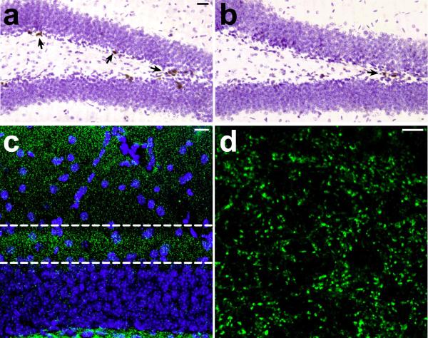
Figure 3. AraC treatment reduces cell proliferation, without altering synaptic marker immunoreactivity.
(a–b), Following a single injection of 300mg kg–1 BrdU with a 24 hour survival period, we observed a significant reduction in the number of labeled cells in wildtype mice that had been infused with the antimitotic drug AraC for ten to fourteen days. The micrograph in (a) shows the dentate gyrus of a mouse infused with vehicle, and the micrograph in (b) shows the dentate gyrus of a mouse infused with AraC. Arrows indicate labeled cells. (c), We also quantified synaptic marker expression in the inner third of the dentate molecular layer, where the medial perforant path synapses are located. This confocal micrograph shows the anatomical region (outlined) where scans were taken for analysis of synaptophysin labeling, and where electrodes were positioned for electrophysiological recordings in slices. (d), Micrograph taken at the resolution and scale used for analysis of synaptophysin labeling (see Supplementary Methods). We observed no differences in the area or intensity of staining for the synaptic marker synaptophysin, suggesting that AraC treatment does not result in loss of synapses among the larger population of dentate granule neurons.

