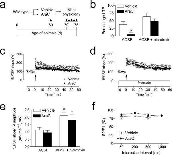
Figure 4. Antimitotic treatment selectively impairs dentate gyrus LTP recorded in the absence of picrotoxin.
(a), Experimental design for studies using minipump delivery of antimitotic drugs. (b), Comparison of the amount of LTP in vehicle– and AraC–infused mice, when recordings were made in the presence or absence of the GABAA antagonist picrotoxin (100μM). Asterisk (*) indicates significance at p < 0.05 following 2 × 2 ANOVA. (c), LTP at medial perforant path synapses in the dentate gyrus is impaired in AraC–infused mice. (d), LTP recordings in the presence of picrotoxin were not influenced by AraC infusion. (e), The relationship between the slope of the dendritic field potential and the amplitude of the axonal fiber volley is influenced by picrotoxin, but not by AraC treatment. (f), Presynaptic paired–pulse depression, measured in plain ACSF, was not altered by treatment with AraC. Error bars = s.e.m.

