
Figure 1. Caveolae integrity is required for Ang II modulating BK channels of rat coronary arterial SMC.
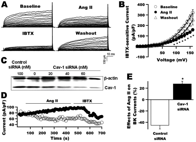
A: Whole-cell K+ currents of rat coronary arterial SMC at baseline, on exposure to 2 μmol/L Ang II and 0.1 μmol/L IBTX and after washout. B: I–V curves of BK currents at baseline, after application of Ang II and washout (n=7). C: Immunoblot shows coronary arterial SMC 48 h after transfection with 0, 20, 40, 60, and 100 nmol/L cav-1 siRNA, and with 100 nmol/L control siRNA. D: Time course of whole-cell K+ currents (HP=-60 mV, TP=+100 mV) before and after the application of 2 μmol/L Ang II and 0.1 μmol/L IBTX. Ang II suppressed the BK currents by about 50% in 100 nmol/L control siRNA treated cells (○), but not in cells treated with 100 nmol/L cav-1 siRNA (●). E: Group results (n=6) were obtained from panel D experiments after the Ang II and IBTX effects have reached steady-state. *: p<0.05 vs. control.