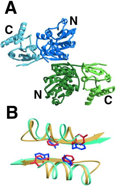
Figure 2.
Dimer Structure. (A) Structure of the MjDEAD dimer found in the asymmetric unit in the crystal with the molecules related by an approximate 2-fold symmetry axis. The individual monomers are shown in blue and green. Two equivalent β-strands (no. 7) are hydrogen bonded, effectively extending the β-sheet to 14 strands. (B) Closeup of the dimer interface of MjDEAD and its superposition with the B:D interface of insulin (PDB code 1trz). Each interface is created by a similar interaction across a roughly 2-fold symmetry axis of two α-helices and two hydrogen-bonded β-strands, depicted as coils and arrows. Insulin is colored yellow with red side chains, and MjDEAD is colored cyan with blue side chains. The arrangement of equivalent aromatic residues on the β-strands (YsF for MjDEAD, FfY for insulin) is shown.

