Figure 6.
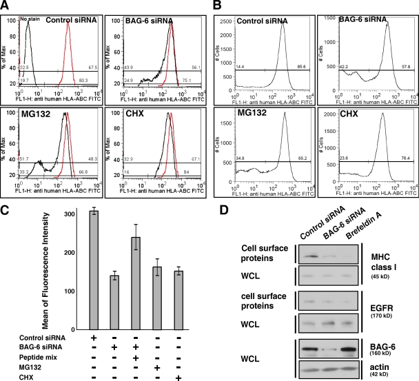
BAG-6 modulates expression of MHC class I molecules on the cell surface. (A and B) Live-cell flow cytometric analysis of HeLa cells with FITC-labeled antibody against MHC class I that recognizes cell surface HLA-ABC antigen. As positive controls, the results of treatment with 10 µM MG132 (A and B) and 10 µg/ml CHX (A) are shown. The data were obtained by linear scale analysis (A) and by log scale analysis (B). The flow cytometric pattern of negative control siRNA is indicated as a red line, and those after treatment with BAG-6 siRNA, MG132, and CHX are indicated with black lines (A). BAG-6 knockdown was performed with two independent duplex siRNAs as described in Materials and methods, and both siRNA gave a similar result. Representatives are results with duplex siRNAs of BAG-6-2 (5′-ATGATGCACATGAACATTC-3′). (C) Quantitative evaluations of the mean of fluorescence intensity of FITC-HLA-ABC on the surface of HeLa cells. Antigenic peptide mixture was pulsed with cells at 50 µM for 16 h. The data shown are the results of at least three independent experiments. (D) Biotin-labeling experiments. Cell surface proteins were biotinylated and affinity purified with avidin beads and blotted with antibody against MHC class I. Knockdown of BAG-6 reduced the expression of MHC class I on the cell surface, whereas the amount of EGF receptor (EGFR) on the cell surface was not affected. Brefeldin A treatment was used as a positive control for general suppression of transport of MHC class I and EGFR to the cell surface. Actin blots for whole-cell lysates (WCL) were used as a loading control for whole cellular proteins.

