
Figure 5.
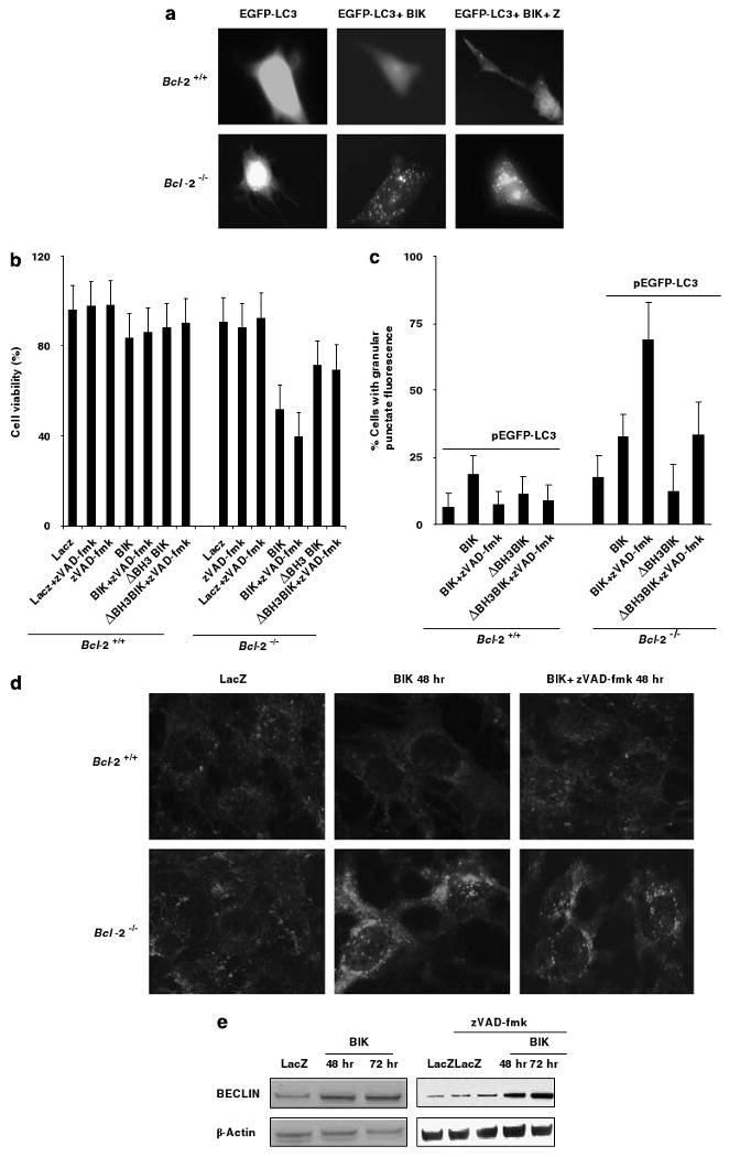
Autophagic features of BIK-induced cell death. (a) Distribution of LC3 in cells transfected with BIK (±zVAD-fmk). (b) Role of BIK BH3 domain on cell death. Cells were infected with Ad-LacZ or Ad-BIK or Ad-BIKΔBH3 (±zVAD-fmk) and viabilities were determined after 48 h; n = 3. (c) Quantification of cells showing punctate distribution of LC3; n = 3. At least 150 cells per sample were examined at random. (d) Immunofluorescence analysis of LC3 distribution. (e) Expression of Beclin-1 in BIK-expressing Bcl-2−/− cells treated with zVAD-fmk.