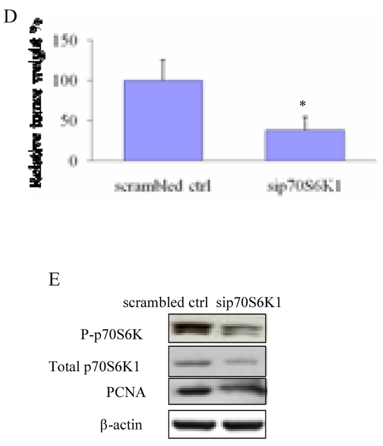
Fig. 3. Sip70S6K1 expression inhibited ovarian tumor angiogenesis and tumor growth in CAM model.
(A) Two million of OVCAR-3 cells expressing sip70S6K1 or scrambled control were collected in serum-free medium and mixed with the equal volume of Matrigel. The mixture or the Matrigel alone was placed on the CAM of 9 days old chicken embryos. Angiogenesis of tumors in the CAM was photographed 5 days after implantation. The experiments were repeated twice with five embryos for each group and representative fields were photographed. (B) The relative blood vessel density was determined by measuring the number of blood vessels in a unit area on Day 5. Results are presented as mean ± SD from replicate experiments, and normalized to results obtained with the CAMs implanted by Matrigel alone. (C) The cells were treated and implanted onto CAM as above. The tumors were harvested after incubation for 9 days. The tumors were photographed and weighed. (D) Tumor weight was obtained from ten tumors in different embryos in each treatment. (E) Parts of tumor tissues from chicken embryos were frozen in liquid nitrogen, and total proteins were extracted and analyzed. Immunoblotting analysis was performed using antibodies against PCNA, phospho-p70S6K1 (Thr421/Ser424), total p70S6K, and β-actin. *, indicates the significant difference (p<0.05).


