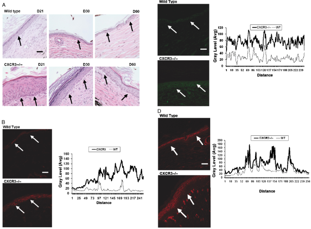
Figure 4.
Delayed in basement membrane remodeling in wounds in mice lacking CXCR3. (A) Representative histological sections of wounds stained with hematoxylin and eosin (H&E) show the immature basement membrane of wounds in CXCR3−/− mice with appearance of gaps between the dermis and epidermis that occur during the fixing and staining process (arrows). (B–D) Immunofluorescence localization of (B) Collagen IV (90 days postwounding), (C) Laminin 5 (60 days postwounding), and (D) Collagen VII (60 days postwounding) shows increased expression in the wounds in CXCR3−/− mice in comparison to those in wild-type (WT) mice. The basement membrane is late in the healing phase of WT mice and resembles a quiescent epidermis basement membrane junction of unwounded skin (not shown). B–D Quantitative measurements of the fluorescence staining in the epidermis portion of the tissues were measured by scanning fluorescence levels across the wounds (demarcated by arrows). All images are representative of six mice. The photomicrographs measure 300 µm on each side; original magnification, ×400. The white bar in the photomicrographs measures 50 microns. CXCR3, cysteine-X amino acid-cysteine receptor 3.

