Figure 1.
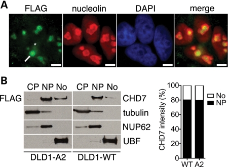
CHD7 localizes to the nucleoplasm and nucleolus. (A) Immunofluorescent staining of DLD1-A2 cells with FLAG and nucleolin antibodies. The arrow indicates nucleolar localization of CHD7, whereas the asterisk marks nucleoplasmic CHD7. Scale bar = 4 µm. (B) Western blot analysis of subcellular fractions from DLD1-A2 and DLD1-WT cells. The purity of cytoplasmic (CP), nucleoplasmic (NP) and nucleolar (No) fractions was assessed by blotting for tubulin, NUP62 and UBF, respectively. Densitometric quantification of CHD7 in the nucleoplasmic and nucleolar fractions is also shown and expressed as a percentage of total CHD7 signal.

