Figure 2.
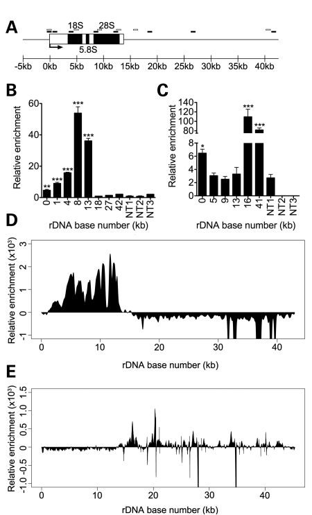
CHD7 binds to rDNA. (A) Schematic representation of a mammalian rDNA repeat. Approximate locations of qPCR primers for human ChIP are denoted by black boxes and mouse ChIP by open boxes. (B) ChIP–PCR analysis of FLAG-CHD7 binding to the human rDNA locus. (C) ChIP–PCR analysis of CHD7 binding to the mouse rDNA locus. For ChIP–PCR, non-target regions are included as controls for specificity of CHD7 enrichment. Error bars represent the mean + SD for triplicates. ChIP-seq of FLAG-CHD7 at human rDNA in DLD1-A2 cells (D) and mouse rDNA in mES cells (E) recapitulates the binding patterns of CHD7 seen by ChIP–PCR. *P < 0.05; **P < 0.01; ***P < 0.001 by t-test versus non-target regions.

