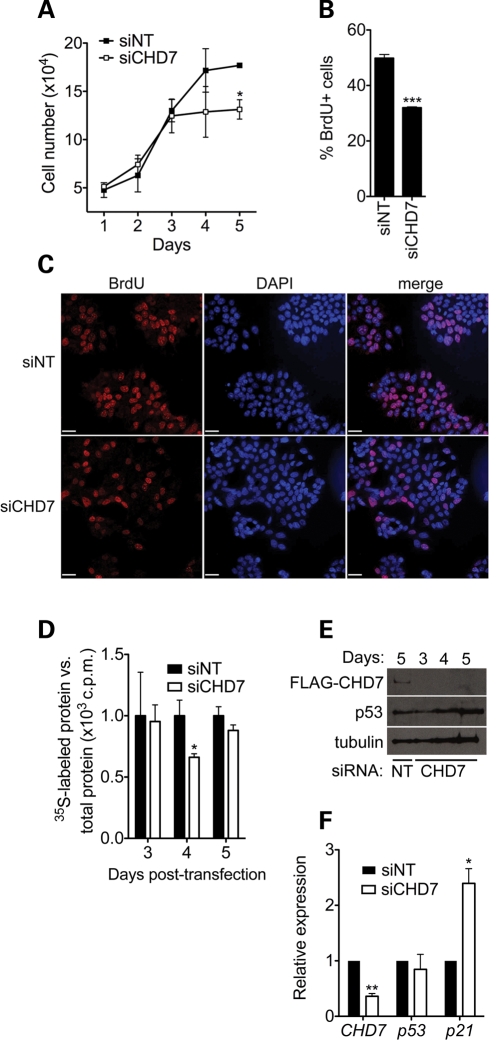
Figure 4.
Loss of CHD7 impairs cell proliferation and protein synthesis. (A) Cell counting assay performed in DLD1-A2 cells treated with the indicated siRNA. Error bars represent mean + SD (n = 3). (B) Quantification of BrdU labeling 5 days post-transfection in DLD1-A2 cells treated with control or CHD7 siRNA. Error bars represent mean + SEM (n = 2–3). (C) Representative image of BrdU labeling in control and BrdU labeling in DLD1-A2 cells showing a qualitative reduction in labeling in CHD7 siRNA-treated cells. Fields are approximately matched for cell number (160–180 cells/field). Scale bar = 32 µm. (D) Measurement of global protein synthesis in DLD1-A2 cells after CHD7 knockdown by [35S]methionine radiolabeling at the indicated time points after siRNA transfection. Scintillation counts were normalized to total protein. (E) Western blots of FLAG-CHD7 and p53 in DLD1-A2 cells. These blots indicate that the CHD7 siRNA is effective up to 5 days post-transfection and that p53 protein levels are not altered. (F) qRT–PCR analysis of CHD7, p53 and p21 transcript levels in DLD1-A2 cells 4 days post-transfection with siRNA (n = 4). *P < 0.05; **P < 0.01; ***P < 0.001 by t-test.

