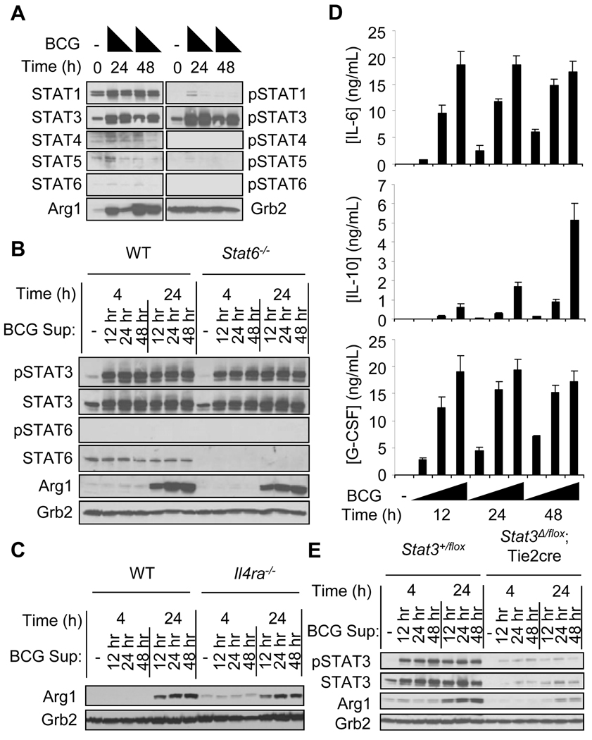
Fig. 2.
The activation of STAT3, but not STAT6, is critical for the induction of Arg1 expression after infection with BCG. (A) BMDMs from wild-type mice were left untreated, or were infected with BCG (at MOIs of 100 or 10). Whole-cell lysates were analyzed by Western blotting for the indicated proteins. Data shown are from one experiment with pooled BMDMs from six mice. Culture supernatants from wild-type BMDMs infected with BCG for 12, 24, and 48 hours were used to stimulate BMDMs from wild-type and Stat6−/− mice (B), and BMDMs from wild-type and Il4ra−/− mice (C) for 4 and 24 hours. Whole-cell lysates were analyzed by Western blotting for the indicated proteins. Data shown are from one experiment with three mice (B) and from one experiment with one mouse (C). (D) IL-6, IL-10, and G-CSF were detected by Luminex in supernatants from BMDMs from wild-type mice infected with BCG at MOIs of 100, 10, and 1. (E) BMDMs from Stat3+/flox mice or Stat3Δ/flox;Tie2cre mice were stimulated with supernatants from BMDMs from wild-type mice infected with BCG at an MOI of 100. Whole-cell lysates were analyzed by Western blotting (n=1 based on deletion efficiency of STAT3). Data shown in D and E are from two experiments, except for the G-CSF data in (D) which are from one experiment.

