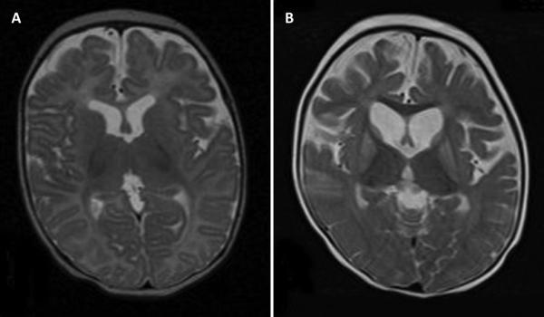
Figure 2.

Brain imaging
Legend: A. Normal brain MRI at age two months. Axial T2 image at the level of the foramen of Monroe shows normal signal intensity and volume of bilateral basal ganglia, normal ventricular size and normal cerebral volume. B. Abnormal brain MRI at age 14 months. Axial T2 image at the level of the foramen of Monroe shows symmetric abnormal increased signal and volume loss in bilateral basal ganglia. There is cortical and subcortical volume loss with enlargement of the lateral and third ventricles and enlarged cerebral sulci. Myelination is normal.
