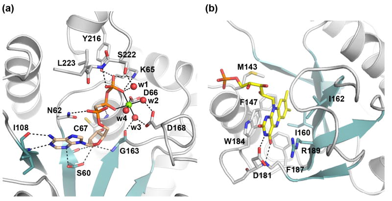
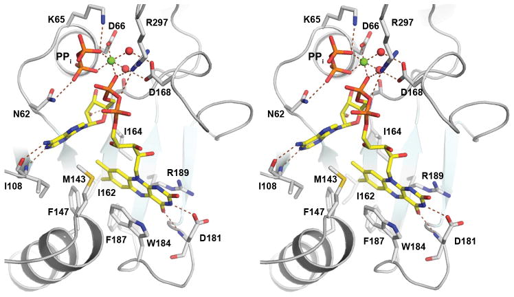
Figure 4. Substrate and product binding in CgFMNAT.
(a) Details of the Mg2+ATP-binding site. Mg2+AMPCPP in the catalytically relevant conformer I is shown. Protein residues interacting with bound subtrates are shown as sticks. The Mg2+ ion is shown as a green sphere and water ligands are shown as red spheres. Hydrogen bonds are shown as dash lines and metal ligands are indicated with solid lines.
(b) Details of FMN binding site.
(c) Stereo view of FAD and PPi product binding site.


