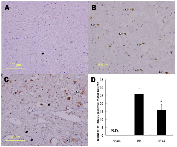
Figure 2.
Micrographs of TUNEL staining and quantification of apoptotic motor neurons 48 h after reperfusion (×400). (A) No TUNEL-positive neurons could be detected in sham group. (B) Many TUNEL-positive neuron with intense nucleus staining were visible in I/R group. (C) Only a small number of positively stained neurons were observed in the HSYA-treatment group. The arrows indicate normal motor neurons, and the arrowheads indicate TUNEL-positive motor neurons. (D) Quantitative analysis of the number of TUNEL-positive cells in the anterior horn of spinal cord of lumbar segments in three groups. * P < 0.05 versus I/R group. N.D. = not detected. The arrows indicate normal motor neurons, and the pointers indicate TUNEL-positive motor neurons.

