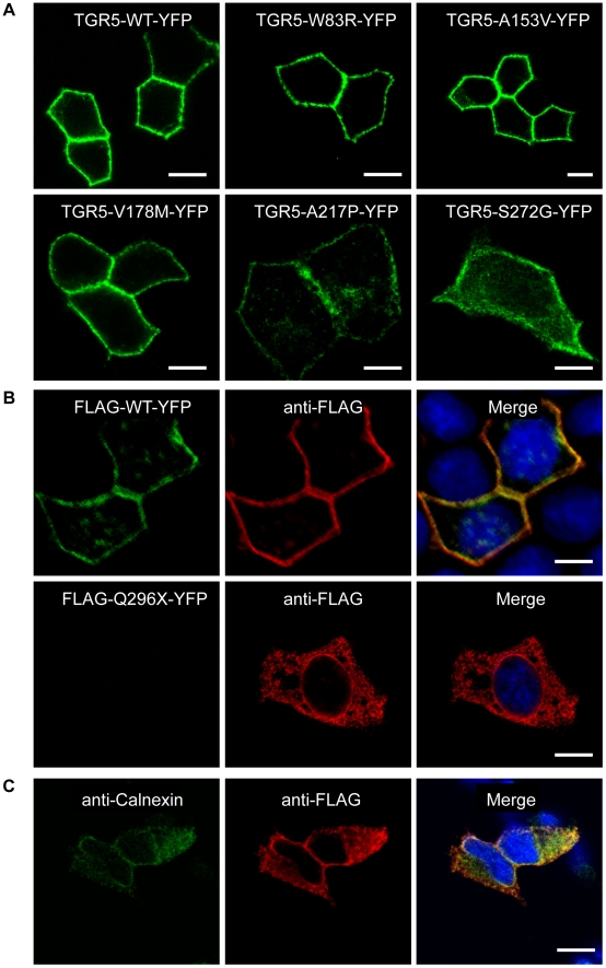
Figure 3. Localization of TGR5 by confocal microscopy.
In Panel A, polarized MDCK cells were transiently transfected with the different TGR5-YFP variants including wild-type (TGR5-WT-YFP). All variants except TGR5-Q296X-YFP reached the plasma membrane, however TGR5-A217P-YFP and TGR5-S272G-YFP were also present in some intracellular vesicles. In Panel B, MDCK cells were transiently transfected with FLAG-TGR5-YFP constructs (wild-type and the Q296X mutant). The FLAG-tag was made visible using an anti-FLAG-M2 antibody (in red) and the yellow coloring in the overlay image demonstrates that the FLAG antibody completely binds to the FLAG-TGR5-YFP wild-type protein both in the plasma membrane as well as in intracellular vesicles (upper row). Introduction of the mutation Q296X leads to a premature stop codon and results in a truncated FLAG-Q296X-YFP protein as demonstrated by the absence of the YFP-fluorescence, but by using the anti-FLAG antibody the truncated protein could be detected (lower row). In Panel C, HEK293 cells were transiently transfected with the Q296X mutant of FLAG-TGR5-YFP (for the remaining variants, see Figure S5). The truncated protein, stained with the anti-FLAG antibody, was almost completely retained in the endoplasmatic reticulum, as demonstrated by the colocalization with the endoplasmatic reticulum marker calnexin. Nuclei were stained with Hoechst. Bars = 10 µm.

