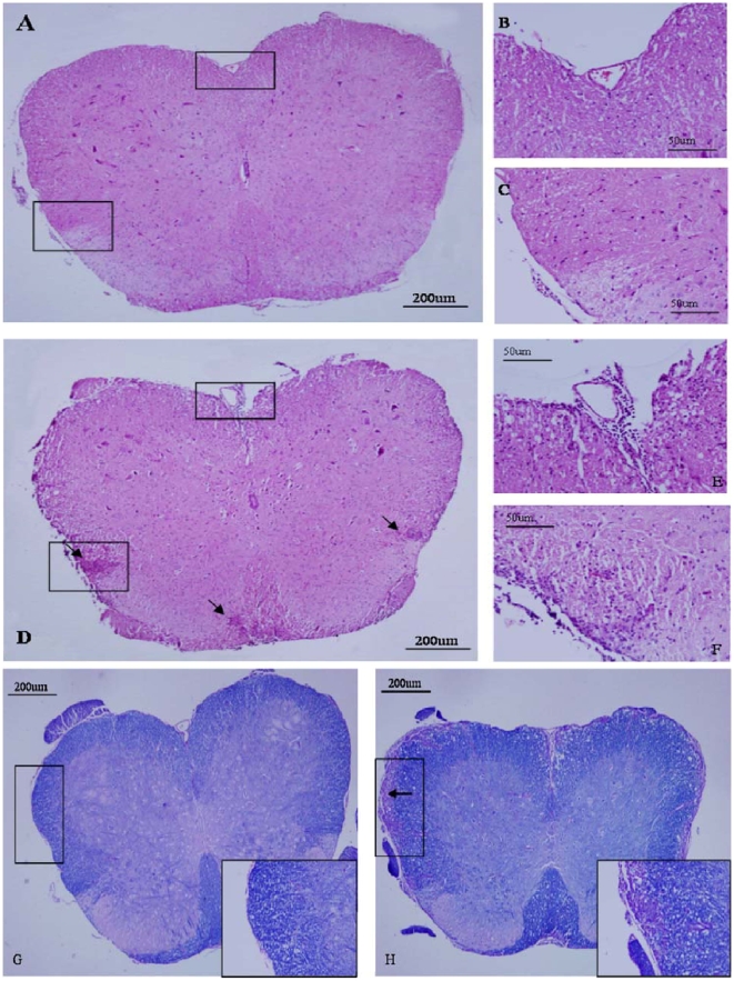
Figure 4. Attenuation of the progression of inflammation and tissue injury in the CNS of mice that received PTx icv.
Pathological examination of spinal cord sections from EAE +PTx icv and EAE mice were performed at 7, 14, and 23 days post EAE induction to evaluate CNS inflammation, demyelination and axonal damage. In EAE +PTx icv mice, the number of immune-cell infiltrates (H&E staining, Fig. 4A–C) and demyelination (Luxol fast blue staining, Fig. 4G) were both significantly reduced at day 14 and 23 post EAE induction. Representative day 14 images of H&E staining (A–F) and LFB/PAS staining (G, H). B and C were inserts in A; E and F were inserts in D. Original magnification ×40 in A, D, G and H; ×200 in B, C, E, F, and inserts in G and H.

