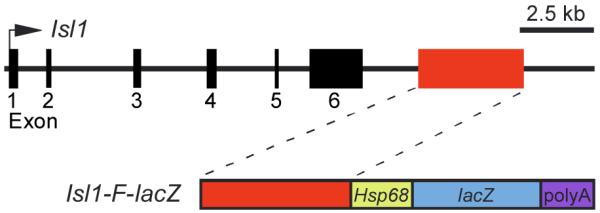
Fig. 1.

Schematic representation of part of the Isl1 locus and the Isl1-F transgene. The top line represents an approximately 18-kb region of the mouse Isl1 locus, including its six exons (black boxes). Exon 1 is the first coding exon. Exon 6 contains the translational stop codon and 3′ untranslated region. The Isl1-F enhancer is represented as a red box in the upper diagram. The lower schematic depicts the transgene construct Isl1-F-lacZ, which contains the 3642-bp Isl1-F fragment in the transgenic reporter plasmid HSP68-lacZ.
