Figure 7.
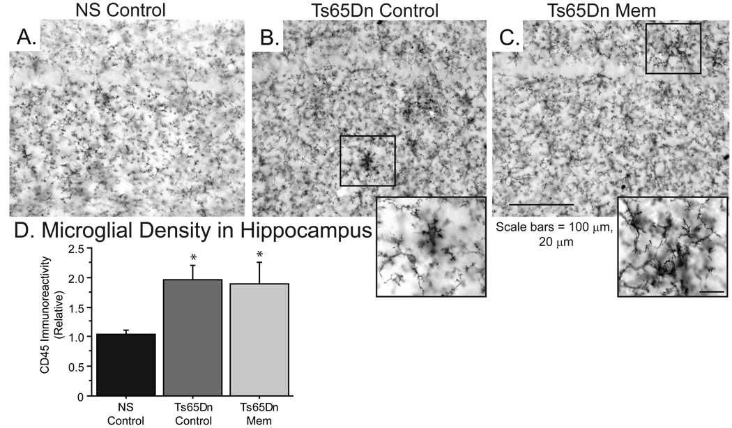
Microglial activity was not reduced in the hippocampus of Ts65Dn mice following memantine treatment. A–C, CD45-positive microglia in the CA1 of the hippocampus exhibit lower immunoreactivity indicative of a “resting” state in NS Control mice (A). Ts65Dn control (B) and memantine-treated (C) mice show larger microglia with greater staining density, consistent with an increased microglial activity (scale bars = 100 µm, 20 µm for inset). D, Microglial immunoreactivity in the hippocampus is elevated in Ts65Dn mice, and is not affected by memantine-treatment (*p<0.05; Immunoreactivity normalized to background; mean ± SEM).

