
Figure 7.
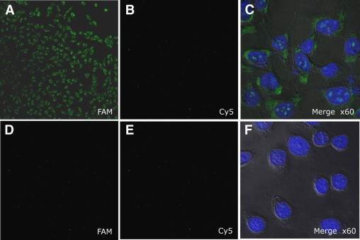
Fluorescence microscope imaging of cells using T790M specific molecular beacons: Target T790M-MB was labeled with FAM fluorophore and control-MB (scrambled molecular beacon) was conjugated with Cy5 fluorophore. Two types of molecular beacons were incubated with H1975 cancer cells that had the secondary mutation in EGFR mRNA (top row) and with the PC9 cancer cells without the secondary mutation (bottom row). FAM: fluorescent images filtered by 550 to 560 nm wavelength band pass (A and D), and Cy 5 fluorescence images were obtained filtered by 650 nm wave length long pass (B and E). DAPI (4’,6-diamidino-2-phenylindole) fluorescent image after DAPI staining; all images were magnified ×10, except Merge ×60, which were magnified ×60 (C and F).