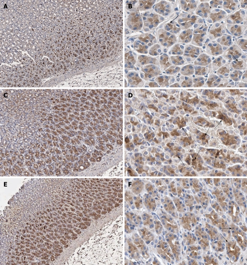
Figure 3.
Immunohistochemical staining of nuclear factor-κB p65 antibody in representative tissue specimens. A, B: Control rats; C, D: Helicobacter pylori (H. pylori)-infected rats; E, F: H. pylori-infected rats supplemented with 200 mg/kg curcumin. Nuclear counterstaining was performed with hematoxylin. The examples of immunoreactive cells are those with dark brown stain in their nuclei (arrows). Images were obtained at × 100 (A, C and E) and × 400 (B, D and F).

