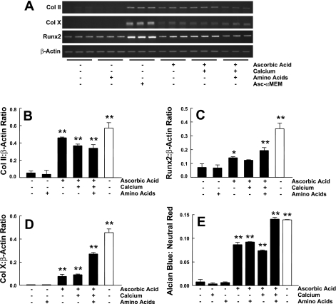
Fig. 1.
ATDC5 cell differentiation in various medium conditions. A: ATDC5 cells were incubated with DMEM-F-12 supplemented with 90 mg/ml calcium, nonessential amino acids, or 50 μg/ml ascorbic acid alone or in combination. Cells were also maintained in standard α-MEM that contains ascorbic acid (Asc-α-MEM). Three markers of differentiation, collagen (Col) II, Runx2, and Col X, and a control gene, β-actin, were assessed using semiquantitative RT-PCR. Triplicate samples were analyzed for each condition. Quantification of these results, calculated as the ratio of Col II (B), Runx2 (C), and Col X (D) to β-actin expression, is shown as means ± 1 SD for triplicate determinations. E: proteoglycan accumulation was assessed using Alcian blue and normalized for cell content using neutral red staining. The dyes were extracted, and absorbance was determined. The ratio of Alcian blue to neutral red was calculated (n = 3 for each condition). Filled bars, DMEM-F-12; open bars, Asc-α-MEM. *P < 0.05 vs. DMEM-F-12 alone; **P < 0.01 vs. DMEM-F-12 alone, as determined by ANOVA. A 2nd replicate experiment gave similar results.

