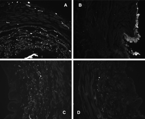
Fig. 6.
Immunofluorescence staining in control (A and C) and VD (B and D) proximal urethras for PGP 9.5 (A and B) and tyrosine hydroxylase (C and D). Images are partial cross sections of the urethra acquired at ×20 magnification. Positive staining is indicated by white areas in the grayscale images. Quantitative measurements of the staining are provided in Table 2.

