Fig. 5.
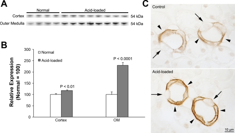
Rhcg expression in IC-Rhcg-KO mice fed a normal and acid diet. A: immunoblot analysis of Rhcg expression in the cortex and outer medulla. Rhcg protein expression in IC-Rhbg-KO mice is increased by acid loading. B: quantification of Rhcg protein expression. Cortical and outer medullary Rhcg expression is significantly greater in acid-loaded than in normal diet IC-Rhcg-KO mice. C: Rhcg immunolabel in the OMCD of normal and acid-loaded diet IC-Rhcg-KO mice. Acid loading increases Rhcg immunolabel in OMCD principal cells (arrowheads) compared with IC-Rhcg-KO mice fed a normal diet. Intercalated cells (arrows) do not express significant Rhcg immunolabel. Values are means ± SE; n = 4, normal diet and n = 8, acid-loaded diet.

