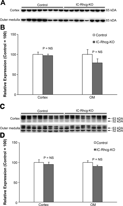
Fig. 9.
PEPCK and PDG expression in acid-loaded IC-Rhcg-KO and C kidneys. A: immunoblot analysis of PEPCK expression in the cortex and OM of acid-loaded IC-Rhcg-KO and C mice. B: quantification of PEPCK protein expression. There was no significant difference in PEPCK expression between acid-loaded C and IC-Rhcg-KO mice in either the cortex or OM. C: immunoblot analysis of PDG expression in the cortex and OM of acid-loaded IC-Rhcg-KO and C mice. D: quantification of PDG protein expression. There was no significant difference in PDG expression between acid-loaded C and IC-Rhcg-KO mice in either the cortex or OM. Expression normalized to mean expression was equal to 100 in control mice in each region for each protein. Values are means ± SE; n = 8/group.

