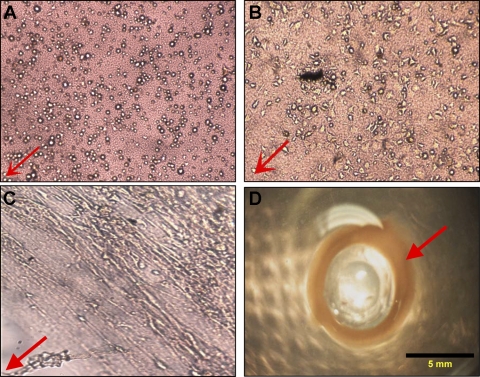
Fig. 1.
Red arrow indicates the direction of the Sylgard post, toward the center of the 35-mm dish. A: ×10 optical image of mouse internal anal sphincter (IAS) cells in the fibrin gels, 15 min after they were seeded. B: ×10 optical image of mouse IAS cells extending in smooth muscle morphology, 4 h postplating on a fibrin gel. C: ×10 optical image showing concentric alignment of cells, 2 days after initial cell seeding, and compaction of the loose fibrin gel with cells and endogenous ECM. D: fully formed bioengineered mouse IAS ring (red arrow), thickened around the Sylgard post. The ring is 2 mm wide and thick, with an inner luminal diameter of 5 mm.

