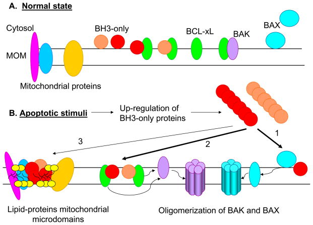
Figure 5.
Models for the apoptotic activity of BH3-only proteins. A. In normal state, BH3-only proteins are present in very low levels in the cytosol or in mitochondrial membrane, most likely in complex with anti-apoptotic BCL-2 family proteins. BAK is shown to be located in mitochondrial membrane in inactive complexes with BH1-4 anti-apoptotic proteins, where as BAX is shown to be in the cytosol or loosely attached to the mitochondrial membrane. B. Different apoptotic stimuli induce significant up-regulation of the BH3-only proteins through transcriptional and post-transcriptional mechanisms. BH3-only proteins then transmit the apoptotic signals to BAX and BAK proteins either through “direct binding/and activation” (1), or “neutralization of BH1-4 proteins and displacement of BAK/BAX” (2). In addition, BH3-only proteins is also shown to induce mitochondrial membrane remodeling/formation of altered microdomains through mitochondrial outer membrane (MOM) insertion and interaction with membrane lipids (e.g., cardiolipin – yellow heads) and/or with other mitochondrial proteins (3). Two classes of BH3-only proteins (that bind with multi domain anti-apoptotic and pro-apoptotic proteins or only with anti-apoptotic proteins) are indicated in red and orange.

