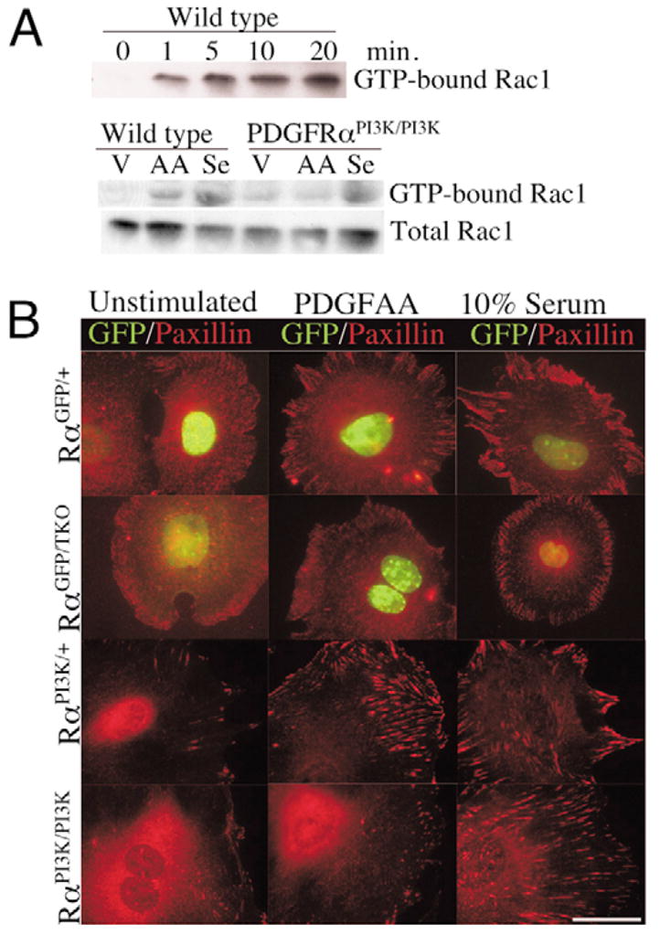
Fig. 6. Effects of PDGFRαPI3K/PI3K mutation on Rac1 activation and paxillin localization.

(A) Western blot for GTP-bound Rac1 and total Rac1 in control and PDGFRαPI3K/PI3K cells. Upper blot demonstrates the time course of Rac1 activation in control cells. Lower blot illustrates the failure of PDGFRαPI3K/PI3K cells to stimulate Rac1 activation when stimulated with PDGFAA. Cells were stimulated for 10 minutes. Results are representative of three independent experiments.
(B) Immunocytochemistry for paxillin localization in control and PDGFRα mutant cells. Genotypes are indicated on the left. Cells were plated for two hours, starved for 24 hours, and then stimulated for 30 minutes with the indicated stimulation. Images are representative of five independent experiments and represent the majority of cells within the culture for each stimulation. Scale bar: 50 μm.
