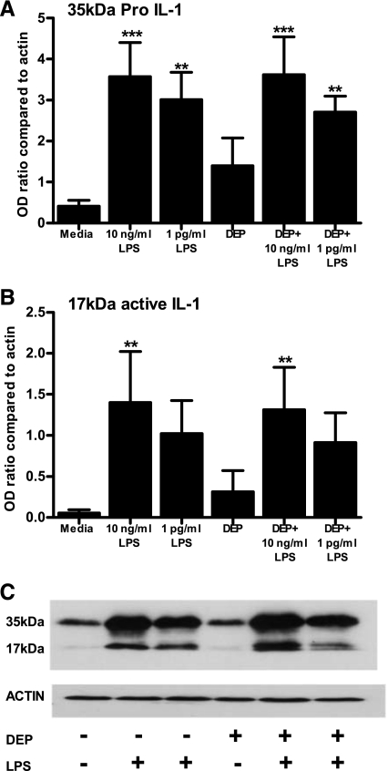
Fig. 6.
A and B: LPS-induced intracellular IL-1 production is maintained in monocytes stimulated in the presence of DEP. Five-hundred thousand monocytes were stimulated in 12-well plates with the indicated agonists for 6 h. Cell lysates were analyzed for pro- and active IL-1β or actin expression by Western blot. Quantitative signals were derived by densitometric analysis using NIH image 1.62, and data displayed as ratio compared with actin loading control. Data shown are means ± SE of n = 4 replicates, each experiment being performed with freshly prepared monocytes from different donors. A representative blot is shown in C. Significant differences compared with unstimulated monocytes are indicated by **P < 0.01 and ***P < 0.001 as measured by 1-way ANOVA and Tukey's posttest.

