Fig. 4.
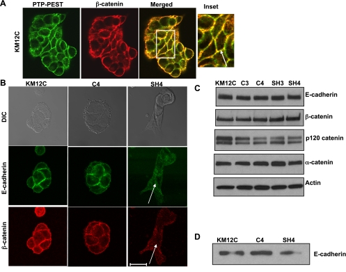
PTP-PEST localizes in cell-cell junctions and affects junction integrity. A: localization of PTP-PEST in KM12C colon carcinoma cells. Confocal image of PTP-PEST (green) and β-catenin (red) staining shows that the two proteins colocalize in adherens junctions (merged) in KM12C cells. Scale bar = 20 μm. B: comparison of C4- and SH4-transfected KM12C cells shows that E-cadherin and β-catenin staining is not detectable in adherens junctions in the SH3 and SH4 knockdown cells (see arrows). Scale bar = 20 μm. C: total protein expression of E-cadherin, β-catenin, p120 catenin, and α-catenin is not altered by PTP-PEST stable knockdown. Actin is shown as a loading control. D: surface expression of E-cadherin is comparable in parental KM12C, C4, and SH4 cells.

