Fig. 1.
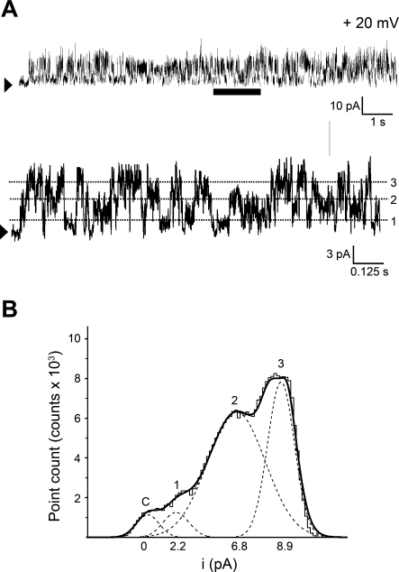
Reconstitution of rat cerebral artery myocyte sarcoplasmic reticulum (SR) membrane protein into lipid bilayers renders unitary current events that correspond to at least 3 conductance phenotypes. A: gap-free channel current recording using Cs+ as permeant ion after SR protein reconstitution into 1-palmitoyl-2-oleoyl-sn-glycero-3-phosphoethanolamine (POPE), 1-palmitoyl-2-oleoyl-sn-glycero-3-phospho-l-serine (POPS), and 1-palmitoyl-2-oleoyl-sn-glycero-3-phosphocholine (POPC) (5:3:2, wt/wt) planar bilayers. This bilayer reconstitution system was used in each of the studies shown in Figs. 2–7, 10, and 11 and in Supplemental Figs. S1 and S2. An arrowhead indicates the baseline (nonconducting channel level); voltage (V) = +20 mV. At the bottom of A, a time-expanded trace from the interval underscored with a horizontal bar is provided. The dotted lines highlight the 3 most frequent unitary current amplitudes (i). Data were low-passed at 1 and digitized at 10 kHz. B: all-points amplitude histogram obtained from the time-expanded interval. The 4 modes correspond to the channel closed state(s), and current levels 1, 2 and 3, which are defined by unitary conductances of 110 ± 8, 334 ± 15, and 441 ± 27 pS, respectively (n = 4–9).

