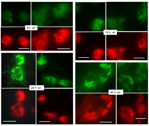
Fig. 1.
The expression of ezrin-wild-type (WT)-yellow fluorescent protein (YFP) in parietal cells increases with time after infection and is distributed primarily to incorporated apical membrane vacuoles. Primary parietal cell cultures were infected with recombinant adenovirus (rAd)-ezrin-WT-YFP construct immediately after plating and subjected to fixation for immunocytochemistry at sequential times afterward, as indicated. Primary antibody against green fluorescent protein (GFP) was used to detect ezrin-YFP (green). Cells were also immunostained for H-K-ATPase (red), which shows a tendency to be redistributed from a primarily general cytoplasmic locale to one more associated with apical vacuoles at the later times of culture. Bar markers = 20 μm.

