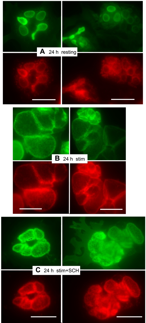
Fig. 2.
Cells expressing ezrin-WT-YFP for 24 h functionally respond to stimulants of acid secretion in a predictable manner. Antibody staining was the same as in Fig. 1 (ezrin-WT-YFP, green; H-K-ATPase, red). A: resting parietal cells show distribution similar to 24-h controls in Fig. 1. B: stimulation with histamine and IBMX effects a large swelling of vacuoles, indicative of HCl accumulation. C: in cells treated with 1 μM proton pump inhibitor SCH28080, as well as secretory stimulants, vacuolar swelling is very much reduced but ezrin-WT-YFP and H-K-ATPase almost entirely colocalize into the apparently thickened walls of apical membrane vacuoles. Bar markers = 20 μm.

